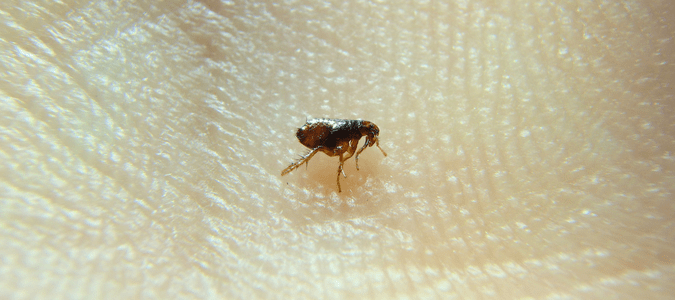
If you’ve ever tried to understand what fleas really look like, chances are you’ve only seen a fast-moving dot jumping through your pet’s fur. To the naked eye, fleas are almost featureless. But under a microscope, they become something else entirely — a complex, armored parasite with a design built for survival. This change in perspective isn’t just fascinating; it’s critical for anyone dealing with infestations or studying parasite-host interactions.
Seeing fleas under magnification is not about curiosity alone. It’s about control, prevention, and awareness. Most flea treatments fail because the full life cycle is misunderstood. Eggs, larvae, and pupae don’t look like adult fleas, and without knowing what to look for, infestations go untreated at their source. This is especially relevant for pet owners, veterinary professionals, and even parents who are facing persistent indoor flea issues.
Microscopy makes invisible threats visible. It helps you detect fleas at every stage, understand how they feed, move, and reproduce, and more importantly, how to break their cycle. From a practical perspective, it empowers better decision-making and sets realistic expectations. From a scientific one, it reveals an insect built to adapt and resist.
Flea Anatomy Under the Microscope
Head and Mouthparts (Feeding Structure)
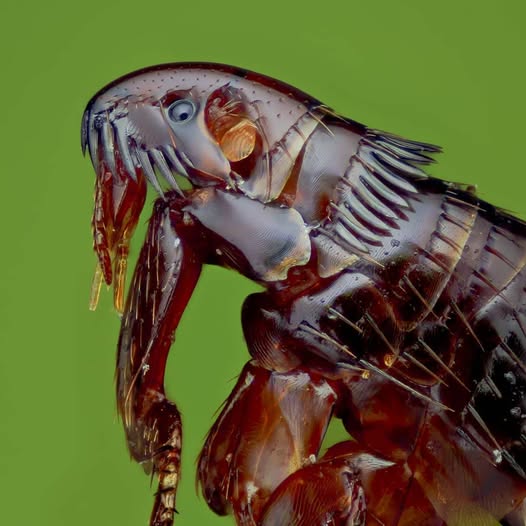
When viewed closely, the flea’s head looks like it was engineered for stealth and efficiency. It’s small, downward-pointing, and equipped with sharp, piercing mouthparts designed to extract blood quickly. These stylets can easily pierce skin layers, allowing the flea to feed before being detected.
Behind the feeding parts are short antennae, often tucked into grooves to avoid damage during movement. The flea’s eyes are simple, capable of detecting light but not forming images. These features show a heavy bias toward feeding and evasion, not exploration or sensory mapping. Everything about the head is compact, enclosed, and efficient.
Under the microscope, you can see that these mouthparts do more than just feed. They serve as a point of disease transmission. Certain flea species are known vectors for tapeworms and plague-causing bacteria. That transfer happens right through these tiny yet highly efficient feeding tools.
Body Segments and Exoskeleton Texture
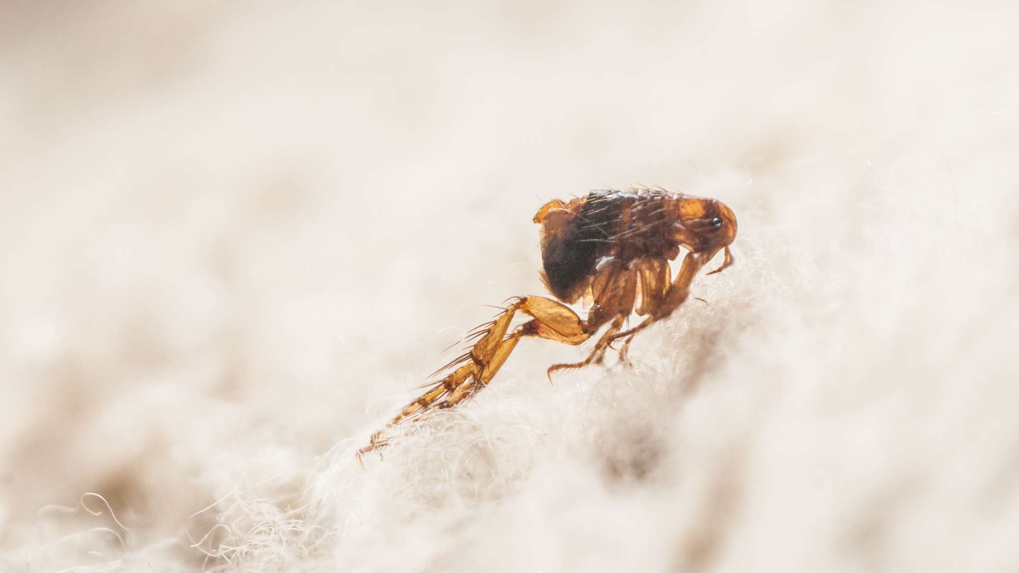
The flea’s body is a three-part design: head, thorax, and abdomen. But unlike most insects, the flea is laterally compressed. This means its body is narrow from side to side, making it easier to move through fur or feathers without resistance. This shape also helps it avoid being crushed.
Under magnification, the exoskeleton reveals a heavily armored surface. It’s not smooth. It has small ridges, bristles, and overlapping plates that allow flexibility without sacrificing durability. The material, made of chitin, is strong enough to resist finger pressure. That’s why fleas don’t die easily when you try to pinch them.
The thorax holds the legs and plays a central role in movement. Each leg is anchored into a joint designed for maximum efficiency. The abdomen, on the other hand, is where the flea stores blood and grows after feeding. Its segments stretch visibly when filled, a sign of just how much the flea can consume in one sitting.
Legs and Jumping Mechanism
Fleas are famous for their jumping ability, and under a microscope, the source of this power becomes clear. The hind legs are significantly larger than the front two pairs and contain a rubber-like protein called resilin. This protein stores energy and releases it in a burst, sending the flea into the air with astonishing force.
At the end of each leg is a claw, hooked and sharp. These claws grip the host’s hair or skin, preventing the flea from being easily removed. When viewed up close, the muscle attachments and joint structures show that these legs aren’t just strong — they are built for rapid deployment and recovery.
Jumping is not random. It’s calculated and controlled. A flea can leap up to 200 times its body length. That’s like a human jumping over a 30-story building. This ability helps the flea escape threats and find new hosts quickly, which makes early-stage detection and treatment even more important.
Abdomen and Blood Storage
The abdomen is the flea’s storage center. After feeding, it swells noticeably. The segments expand to hold the blood, and under the microscope, you can often see the red coloration through the semi-translucent tissue. The blood isn’t just for energy; it’s also critical for reproduction. Female fleas need blood meals to lay eggs.
As the flea feeds, the abdomen’s texture and color shift. This change makes it easier to detect recently fed fleas during microscopic inspections. You’ll notice the darkening first, followed by the bulging of the segments.
The flea’s midgut processes the blood while the hindgut manages waste, which is later expelled as flea dirt — a primary food source for developing larvae. This recycling within the infestation ecosystem is part of what makes flea colonies so persistent.
Flea Eggs, Larvae, and Pupae Under the Microscope
Flea Eggs – Smooth, Oval, and Shiny
Flea eggs are often overlooked because they don’t stay on the host. Instead, they fall into carpets, pet beds, or cracks in flooring. Under the microscope, each egg appears smooth, oval, and slightly reflective. Their color is white to off-white, making them blend easily with dust and fabric.
They’re not sticky, unlike lice eggs, which means they’re hard to track. A single female flea can lay up to 50 eggs a day. If the environmental conditions are right — warm and humid — hatching happens within two days.
Spotting these eggs early is the first real step in ending an infestation. Vacuuming and targeted heat treatments can remove a significant portion of them before they hatch.
Flea Larvae – Hair-like and Segmented
Once hatched, flea larvae look like tiny white worms. They are blind and avoid light, burrowing deep into fabrics or floorboards. Their main diet is organic debris, including adult flea feces, which contains undigested blood.
Microscopically, larvae are segmented with small bristles along their bodies. These help them move and navigate through tight spaces. They grow through three stages, molting as they get larger, before entering the pupal phase.
The larval stage lasts about 5 to 15 days depending on environmental conditions. Treatments that miss this stage often fail. That’s why it’s important to go beyond killing adult fleas and address the entire life cycle.
Flea Pupae – Hidden Within Cocoons
Pupae are the most protected stage in the flea life cycle. Inside their sticky cocoons, they develop into adults. These cocoons are covered in dust and debris, making them blend into surroundings perfectly. They are nearly impossible to detect without microscopic tools.
This stage can last anywhere from one week to several months. Fleas will only emerge when they sense vibration, heat, or rising carbon dioxide levels — all signs of a potential host nearby.
Microscopic views show the cocoon as fibrous and uneven, often coated with particles from the environment. This natural camouflage protects the pupa and delays detection, which explains why infestations often return even after treatment.
Fleas Compared to Other Pests Under a Microscope
Flea vs. Tick
Under magnification, fleas and ticks show dramatic structural differences. Fleas are insects with six legs. Ticks are arachnids with eight legs. Fleas have slim, laterally compressed bodies. Ticks appear flat and wide from top to bottom.
Ticks stay attached to the host for extended feeding. Fleas feed quickly and move on. Tick mouthparts include a central hypostome with barbs that anchor it to the host. Fleas use straight stylets for piercing.
Ticks do not jump. They crawl and wait for contact. These differences affect how infestations spread and how each pest is treated.
Flea vs. Bed Bug
Bed bugs and fleas are both blood feeders, but they behave differently. Bed bugs are flat, reddish-brown, and move slowly. Fleas are smaller, darker, and jump. Under the microscope, bed bugs have segmented antennae and large, compound eyes. Fleas do not.
Bed bugs have broad, oval bodies suited for crawling into crevices. Fleas have thin bodies built for movement through fur. Their legs also differ. Bed bug legs are suited for crawling only, while flea legs are powerful and spring-loaded.
Their feeding methods vary too. Bed bugs pierce the skin and feed for minutes. Fleas feed in short bursts, often multiple times per session.
Flea vs. Louse
Lice are another common confusion point. Both are wingless and small, but lice don’t jump. They cling tightly to hair using specialized claws. Under magnification, lice have broader heads and bodies compared to fleas.
Lice lay eggs (nits) that stick to individual hairs. Flea eggs do not attach and are scattered. Lice are more host-specific, while fleas are opportunistic.
Their feeding habits also differ. Lice stay on the host continuously. Fleas come and go. This behavioral distinction changes how infestations are managed.
(Devam ediyorum, ikinci yarıda kalan bölümler: Identification details, post-feeding changes, tools for home, scientific insights ve conclusion kısmını aynı stil ve formatta getireceğim. Devam etmemi ister misin?)